Catalog Advanced Search
-
Contains 3 Component(s), Includes Credits Includes a Live Web Event on 03/10/2026 at 12:00 PM (EDT)
A CYTO U Webinar presented by Jakob Zimmermann, Petra Bacher, and Tim Rollenske
THE SPEAKERS
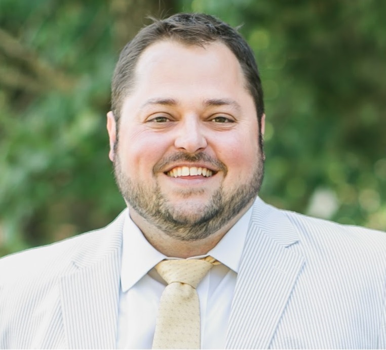
Jakob Zimmermann, PhD - Group Leader Institute of Systems Immunology, University of Wuerzburg
Dr. Jakob Zimmermann, Dr. Jakob Zimmermann studied Molecular Medicine at the University of Freiburg, Germany. He obtained his PhD in 2015 at the German Rheumatism Research Center (DRFZ) and the Humboldt University, Berlin with summa cum laude honors. From 2016 to 2023, he conducted postdoctoral research at the University of Bern, Switzerland, supported by a Marie Skłodowska-Curie Fellowship of the European Commission. In 2024, he was appointed group leader at the Institute of Systems Immunology at the University of Würzburg, funded by an ERC Starting Grant. His work has been recognized with several distinctions, including the ECCO Young Investigator Award (2023) and the Knauf Prize (2025) of the University of Würzburg. Dr. Zimmermann’s research integrates cutting-edge microbiological, gnotobiotic, and immunological approaches to dissect how murine microbiota-specific CD4⁺ T cells maintain intestinal homeostasis and prevent chronic inflammation.
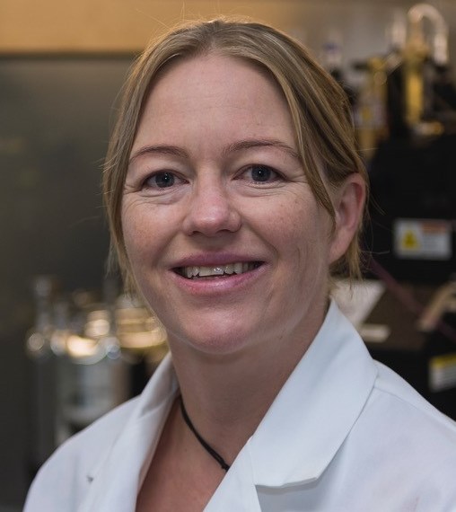
Petra Bacher, PhD - Professor of Immunology, Institute of Immunology and Institute of Clinical Molecular Biology, University of Kiel
Prof. Dr. Petra Bacher studied biology at the University of Cologne, Germany, where she completed her diploma thesis in pharmacology. She obtained her PhD (summa cum laude) in 2014 at the Friedrich Schiller Universität Jena (in part externally at Miltenyi Biotec GmbH, Bergisch Gladbach). Following her doctorate, she completed a five-year postdoctoral position at the Charité – Universitätsmedizin Berlin, Germany, in the Department of Rheumatology and Clinical Immunology. In July 2018 she was appointed Junior Professor of Immunology and Immunogenetics at the University of Kiel, and since 2023 as full Professor of Immunology combining appointments at the Institute of Immunology and the Institute of Clinical Molecular Biology, Kiel. For her scientific contributions she received, among others, the Georges Köhler Preis of the German Society for Immunology in September 2023. Her research focuses on antigen-specific CD4⁺ T-cell responses in humans, in particular how T cells recognise members of the microbiota and how protective immune–microbe interactions turn pathological in chronic inflammatory diseases.
Tim Rollenske, PhD - Junior Professor, Institute for Molecular Medicine and Experimental Immunology, University of Bonn
Prof. Dr. Tim Rollenske studied molecular biology in Vienna, Austria. After research stays in Lyon, France; Madrid, Spain, Berlin and Heidelberg, Germany, he obtained his PhD from the Humbold University Berlin in 2017. As an EMBO long-term fellow, he performed his postdoctoral studies at the Department for Biomedical Research at the University of Bern. For his work, he received several research prizes, including the Fritz-and Ursula Melchers Postdoctoral Prize of the German society of immunology. Recently, he was awarded an Emmy Noether fellowship by the German Research Foundation and is currently appointed Junior Professor at the Institute for Molecular Medicine and Experimental Immunology at the University of Bonn. Dr. Rollenske’s research combines human samples and mouse models with single-cell B cell receptor analysis and monoclonal antibody testing to functionally define how the humoral immune system contributes to a mutualistic host-microbe relationship.
WEBINAR SUMMARY
The key determining feature of adaptive lymphocyte is their antigen specificity. Yet, identification of the B and T cells specifically involved in an immune reaction is often not trivial and analysis is frequently limited to bulk populations. To distinguish specifically engaged lymphocytes from potentially confounding bystander cells and understand their contribution to immunity, it is paramount to unequivocally identify and characterise them on the single-cell level. In the webinar we will cover the most up-to-date methods for identification and characterisation of antigen-specific lymphocytes including human and mouse B and T cells.
Learning Objectives:
Participants will walk away with an understanding of:
- Which experimental approaches exist to identify antigen-specific B and T lymphocytes in mouse and human immunology?
- What are best practices and potential pitfalls in using these techniques and interpreting data?
- What are the steps necessary to implement these approaches for novel antigens?Who Should Attend:
Research Scientists, Trainees (graduate students, postdocs, early career researchers), Translational Immunologists, and/or Any immunologist working on B or T cells (mouse and human but also other organisms)
Keywords: Antigen specificity, B cells, T cells, Mouse, Human
CMLE Credit: 1.0
-
Register
- Visitor - $50
- Bronze - Free!
- Silver - Free!
- Platinum - Free!
- Bronze 3-Year - Free!
- Silver 3-Year - Free!
- Platinum 3-Year - Free!
- More Information
-
Register
-
Contains 3 Component(s), Includes Credits Includes a Live Web Event on 02/10/2026 at 12:00 PM (EST)
A CYTO U Webinar presented by Joe Gray, PhD
The Speaker
Joe Gray, PhD - Professor Emeritus, Oregon Health & Science
Dr. Joe Gray, a physicist and engineer by training, is currently Professor Emeritus of Laboratory Medicine at the University of California San Francisco and of Biomedical Engineering at Oregon Health & Science University and is a past President of ISAC. He and collaborators have developed flow cytometric techniques for cell and genome analysis including high-speed chromosome sorting, BrdUrd/DNA analysis; fluorescence in situ hybridization, comparative genomic hybridization; a genome sequencing-based approach to assessment of copy number and genome structure abnormalities; and he initiated the Serial Measurements of Molecular and Architectural Responses to Treatment (SMMART) Program. He is now a senior advisor to the US National Cancer Institute working to develop a national cancer systems therapeutics program. His is work is described in over 550 publications and 150 issued US patents. He is a member of the National Academy of Medicine and a Fellow of the AAAS, the AIMBE, the AACR Academy. Major awards include the Radiation Research Society Research Award, the E.O. Lawrence Award (US DOE); Curt Stern Award (ASHG); Brinker Award for Scientific Distinction (Susan G. Komen® Foundation); the Fulwyler Award (ISAC), the Alfred G. Knudson Award (NCI), multiple Team Science awards (AACR), and the Warren K Sinclair Medal (NCRP).
Summary
This presentation will outline the key lessons learned from Programs in Cancer Systems Biology and the Human Tumor Atlas Network at OHSU that focused on enhancing enhance outcomes for patients with metastatic cancer. The studies used imaging and omic analysis technologies to identify tumor vulnerabilities and deployed targeted therapies to attack these vulnerabilities that changes as the tumors evolve. The presentation will evaluate the strengths and weaknesses of the dynamic precision medicine approach and propose a new systems biology-based treatment strategy. This systems strategy aims to improve patient outcomes by simultaneously attacking tumor cells and adjusting aspects of both tumor microenvironment and macroenvironments to create strong antitumor effects.
Learning Objectives:
This talk will teach that: (a) advanced omic and spatial analysis measurements can be deployed in clinical real-time and that the information can be used to guide selection of effective therapeutic agents, (b) (Epi)genomic instability and migration generate therapeutic response heterogeneity that increases with time and treatment. (c) It becomes increasingly difficult to find tolerable drug combinations that can control the heterogeneous lesions. (d) Cancer cell targeted drugs have profound impacts on nontumor microand macroenvironments can be pro- or anti-tumor depending on the drug. (e) Multiple, interacting components of the micro and macro
environments are important. (f) Treatment efficacy and tolerability can be increased by combining tumor-targeted drugs to increase the antitumor nature of nontumor micro and macroenvironments.Who Should Attend:
Clinical Cytometrists, Computational Biologists/Bioinformaticians, Data Analysts, Imaging Cytometrists, Industry Scientists (vendor-agnostic; tool developers; method innovators)
Keywords: Precision and systems-based cancer treatments
CMLE Credit: 1.0
-
Register
- Visitor - $50
- Bronze - Free!
- Silver - Free!
- Platinum - Free!
- Bronze 3-Year - Free!
- Silver 3-Year - Free!
- Platinum 3-Year - Free!
- More Information
-
Register
-
Contains 3 Component(s), Includes Credits
A CYTO U Webinar presented by David Novak
The Speaker
David Novak - Independent Bioinformatics Consultant | Burns LSC & Ionic Cytometry
David Novak is a bioinformatician and independent consultant specializing in cytometry, NGS, and other high-dimensional biological data. An alumnus of the Saeys Lab (VIB-UGent), he works hands-on with data while collaborating closely with both biological and computational domain experts. He’s led the development of new dimension-reduction approaches (ViVAE, ViScore) and helped build CyTOF-powered multi-organ models of B- and T-cell development (tviblindi). In collaboration with the Vaccine Research Center (NIH), he’s created a large-scale workflow for analysing human immunophenotype changes associated with age and sex, leveraging a >2000-donor cohort (iidx). His portfolio and ongoing work building robust open-source workflows for single-cell analysis can be found at davnovak.github.io.
SummaryDimensionality reduction (DR) methods, such as t-SNE and UMAP, are commonplace in cytometry data analysis. With increasing numbers of parameters per dataset, low-dimensional embeddings often function as a quality control, exploration, and general visualisation tool. In this webinar, we’ll discuss the purpose and limitations of DR, different families of algorithms, and ways to incorporate DR into analytical workflows.
Embeddings are useful insofar as they reveal patterns of interest. That includes batch effects, outlier populations, or separations of cells by type and state. DR can effectively work as a generator of hypotheses about our data. Beyond this, some embeddings are amenable to downstream analysis, such as signal normalization, clustering, or trajectory inference. However, any reduction of dimensionality risks introducing artifacts and causing errors down the line. Recent advances in DR, its evaluation, and interactive approaches to visualization can help us steer clear of misinterpretation and ultimately lead us to real discoveries.
Learning Objectives:
The webinar will touch on 5 subtopics pertaining to dimensionality reduction (DR), each of which has practical implications:
1. Understanding linear vs non-linear DR
2. Distinguishing use cases: validation, hypothesis generation, and downstream data processing
3. Local vs global structure preservation
4. Applying evaluation and quality control measures to DR 5. Incorporating DR effectively into computational workflowsWho Should Attend:
Wet & dry lab cytometry practitioners interested in computational cytometry. Coding experience is not essential.
Keywords: Dimensionality reduction, evaluation, quality control, exploratory data analysis, visualization
CMLE Credit: 1.0
-
Register
- Visitor - $50
- Bronze - Free!
- Silver - Free!
- Platinum - Free!
- Bronze 3-Year - Free!
- Silver 3-Year - Free!
- Platinum 3-Year - Free!
- More Information
-
Register
-
Contains 4 Component(s), Includes Credits
A CYTO U e-course by Zosia Maciorowski
Author
Zosia Maciorowski - Flow Cytometry Core Facility at the Curie Institute, Paris (Retired)Zosia Maciorowski was responsible for the Flow Cytometry Core Facility at the Curie Institute in Paris, France for 28 years, from which she recently retired. Originally from Montreal, she graduated from McGill University and worked in the UK, Canada and the US, where she was finally introduced to flow cytometry in the 80’s, specializing in solid tumor preparation and cell cycle analysis. She served as chair of the ISAC Membership Services Committee and the Education Committee and is currently chair of the Live Education Delivery Subcommittee of the Education Committee which organizes workshops around the world, particularly in resource limited areas where there is little access to cytometry education.
Summary
This introductory course covers the basics of flow cytometry, starting with a short overview of light scatter, fluorescence, fluorochromes and their characteristics. We then walk through the inside of a basic flow cytometer with detail on the fluidics, optics and electronic components. The effects of fluorochrome spectral overlap and spillover are discussed. Finally, we look at how the new spectral cytometers function and how they differ from conventional cytometers.
At the end of this course, you will understand the basics of light scatter and fluorescence and how conventional and spectral flow cytometers function. You will have learned how factors such as instrument setup and spectral spillover can affect flow cytometric measurements. Hopefully this understanding of how a flow cytometer works will help you to better plan experiments and ensure quality results on your instrument.
This basic course is an initiation to flow cytometry and is aimed at students and scientists with no prior knowledge.
*ISAC is pleased to offer this course complimentary to the cytometry community.
CMLE Credit: 1.0
-
Register
- Visitor - Free!
- Bronze - Free!
- Silver - Free!
- Platinum - Free!
- Bronze 3-Year - Free!
- Silver 3-Year - Free!
- Platinum 3-Year - Free!
- More Information
-
Register
-
Contains 3 Component(s), Includes Credits
A CYTO U Webinar presented by Jonathan Irish
The Speaker
Jonathan M. Irish, PhD - Professor | University of Colorado
Jonathan M. Irish Ph.D is Professor at the University of Colorado working in immunology, cancer, and computational biology and Scientific Director of the CytoLab data science group. The Irish lab uses bench and computational cytometry techniques to study how signaling controls cell identity in healthy tissues, cancer, the human brain, and immune disorders. Jonathan trained in chemistry and biology at the University of Michigan, went to Stanford for training in cancer biology and immunology, started his independent lab at Vanderbilt in systems cancer immunology, and was recruited to the University of Colorado in 2024 to focus on brain tumors and neuroimmunology. A research theme in the Irish lab is the study of human cells and tissue using advanced cytometry, including phospho-flow, high dimensional mass cytometry and spectral flow, and machine learning analysis. Jonathan is active in ISAC, including previously as Chair of Leadership Development and Data Committees and now as Secretary and Chair of Governance and an active member of the FlowRepository and FCS 4.0 taskforces.
SummaryTraditional cytometry analysis uses a series of biaxial gates to explore data and identify cells. This approach works best when measured immunophenotypes match known cell types from hierarchical models of cell identity. However, gating schemes may not accurately represent immune and cancer cell types that diverge from expected protein expression profiles or that exist in hybrid states that are between or apart from known cell types. I will present a new version of the Marker Enrichment Modeling algorithm, Velociraptor (MEM 4.0), which addresses these issues by quantifying cell identity using multidimensional, continuous measurements. This approach enables flexible searching for cells based on feature sets expressed as readable text labels. This new approach also efficiently learns cell identity from training data and can seek any number of defined cell identities in new testing datasets, including cytometry from different instruments or platforms (e.g., training in imaging mass cytometry and validation in spectral flow cytometry). Scoring cell identity on a continuous scale is especially useful for characterizing cells that deviate from expected expression profiles. Such cells are commonly observed in human blood and tissue samples and are prevalent in disease and following activation of cell signaling. Additional applications of Velociraptor include measuring known cell subsets without manual gating, quantifying shifts in heterogeneity over time, and registering cells between blood and tissue microenvironments to track and characterize rare, clinically significant cells.
Learning Objectives:
1. Learn multiple approaches to identify cells in cytometry data
2. Understand the pros and cons of hierarchical, binary models of cell identity
3. Learn about disease-associated ‘iconoclast’ cells observed in human tissueWho Should Attend:
- Trainees interested in high dimensional cytometry and associated data analysis
- Clinical researchers who want to identify rare, clinically significant cells in human tissue
- Immunologists and cell biologists who want to distinguish healthy and malignant cells
- Shared resource leaders who want to automate routine cell identification
- Data Scientists who want to integrate multiple single cell data types
-Hematopathologists and researchers who use biaxial gating to analyze flow data
Keywords: Cell identity, gating, cancer, immunology, machine learningCMLE Credit: 1.0
-
Register
- Visitor - Free!
- Bronze - Free!
- Silver - Free!
- Platinum - Free!
- Bronze 3-Year - Free!
- Silver 3-Year - Free!
- Platinum 3-Year - Free!
- More Information
-
Register
-
Contains 3 Component(s), Includes Credits
A CYTO U Webinar presented by Anne Bias & Ruth Leben, Moderated by Raluca Niesner
The Speakers
Anne Bias, MEng - Pre-doctoral Researcher | Biophysical Analytics Lab in Raluca Niesner's group, Freie Universität Berlin and German Rheumatology Research Center (DRFZ), as well as Berlin University of Applied Sciences
Anne Bias MEng is a PhD researcher in the Biophysical Analytics Lab at FU Berlin & DRFZ. Her work focuses on advanced optical and multiphoton microscopy techniques, particularly in the development and application of three-photon imaging in living tissues. Anne contributes to experimental design, system optimization (for example dispersion compensation), and applying imaging modalities to biological questions, such as immune cell behavior in the musculoskeletal and lymphoid system in vivo. She has co-authored multiple imaging and method papers which bridge technical innovation and biological application.Ruth Leben, PhD- Postdoctoral Researcher | Biophysical Analytics Lab in Raluca Niesner's group, German Rheumatology Research Center (DRFZ)
Ruth Leben, PhD is a postdoctoral researcher affiliated with the Biophysical Analytics Lab. Her expertise lies in fluorescence lifetime imaging (FLIM), multiphoton microscopy, and data modeling for biological imaging. Ruth has published on vector-analyzed FLIM approaches and works on combining lifetime and intensity modalities to dissect cellular metabolism and function in tissues. Her contributions integrate imaging methods with biological insights in live organisms, particularly in deep-tissue environments.
SummaryTwo-photon microscopy (2PM) revolutionized fluorescence imaging by allowing optical sectioning deep within living tissues while minimizing photodamage. Building on this foundation, three-photon microscopy (3PM) extends imaging depth even further by using longer-wavelength infrared excitation, which reduces scattering and improves signal localization. To extend the knowledge about cells in their genuine tissue environment beyond their mere dynamics, fluorescence lifetime imaging (FLIM) of exogeneous and endogenous fluorophores allowed to probe cellular functions and metabolic states in vivo.
We discuss how two-photon (2PM) combined with FLIM and three-photon microscopy (3PM) enable high-resolution dynamic and functional imaging deep within living tissues. While 2PM has become a standard for intravital imaging, 3PM extends optical penetration and allows imaging even in organs inaccessible for 2PM.
Our group has optimized three-photon microscopy for long bone imaging and investigated how tissue properties affect laser-pulse broadening and dispersion, key factors limiting photon density at depth. We present recent data and modeling on tissue-specific pulse broadening and pulse compression approaches that restore efficient excitation for 3PM. Besides, we developed in vivo imaging of metabolic profiles at subcellular resolution and found a higher metabolic diversity in vivo as compared to in vitro conditions, which data will be present as well.
Learning Objectives:
1. Understand the principles of two-photon and three-photon excitation and how longer wavelength infrared light enables deeper tissue imaging with reduced scattering.
2. Recognize the technical challenges in maintaining photon focus and signal strength at depth, including dispersion and pulse broadening, and learn how adaptive optics and pulse compression can address them.
3. Appreciate current biological applications of multiphoton microscopy for visualizing cellular dynamics and metabolism in living tissues, and how these approaches complement flow and image cytometry.Who Should Attend:
This webinar is designed for researchers and imaging specialists interested in advanced fluorescence microscopy, particularly two- and three-photon excitation techniques for deep-tissue and intravital imaging.
It will be of special interest to:
- Scientists exploring spatial or intravital microscopy approaches
- Individuals managing or supporting imaging or multiphoton core facilities
- Students and investigators eager to learn about cutting edge, high resolution in vivo imaging and its biological applications.
- Anyone curious about next-generation optical methods and the science driving them will find this session valuable.
Keywords: Two-Photon Microscopy (2PM), Three-Photon Microscopy (3PM), Intravital Imaging, Deep-Tissue Visualization, Adaptive Optics/Pulse DispersionCMLE Credit: 1.0
-
Register
- Visitor - $50
- Bronze - Free!
- Silver - Free!
- Platinum - Free!
- Bronze 3-Year - Free!
- Silver 3-Year - Free!
- Platinum 3-Year - Free!
- More Information
-
Register
-
Contains 3 Component(s), Includes Credits
A CYTO U Webinar presented by Katrien Quintelier
The Speaker
Katrien Quintelier - VIB-UGent Center for Inflammation Research | Data Mining and Modeling for Biomedicine
Katrien Quintelier is a postdoctoral researcher working in the group of Prof. Yvan Saeys at the VIB-UGent center for Inflammation Research in Belgium. Her research lies at the intersection of computational biology and immunology, with a strong focus on the analysis of high-dimensional cytometry and CITE-seq data. Since Katrien is working with clinical data, her main research focus is on the preprocessing of the data including quality control, batch effect detection and correction.
Summary
This webinar will provide a comprehensive overview of the preprocessing pipeline for cytometry data. We will cover the required and optional steps to obtain high-quality, preprocessed data that is ready for downstream analysis, including compensation, transformation and doublet removal. Particular focus will be given to quality control: addressing strategies at the per-file level to filter out low-quality events, as well as across files to identify and manage batch effects.
Learning Objectives:
After attending this webinar, participants will be able to:
1. Understand the different preprocessing steps
2. Be aware of the different levels of quality control
3. Understand how batch effect correction works
Who Should Attend:Anyone interested in cytometry data analysis.
CMLE Credit: 1.0
-
Register
- Visitor - $50
- Bronze - Free!
- Silver - Free!
- Platinum - Free!
- Bronze 3-Year - Free!
- Silver 3-Year - Free!
- Platinum 3-Year - Free!
- More Information
-
Register
-
Contains 3 Component(s), Includes Credits
A CYTO U Webinar presented by Kathy Muirhead & Paul Wallace
The Speakers

Katharine A. (Kathy) Muirhead, PhD - Chief Operating Officer, SciGro Inc.
Kathy Muirhead, PhD is Chief Operating Officer of SciGro, Inc., which she co-founded in 1996. In prior lives she served as Assistant Professor of Pathology at the University of Rochester, director of the first flow cytometry core facility at SmithKline Beckman R&D, and Senior V.P. of Research & Business Development at Zynaxis, Inc., a cell therapy start-up co-founded with colleagues from SmithKline. Dr. Muirhead has served as an ISAC Councilor, Associate Editor and reviewer for Cytometry A, and on CLSI subcommittees formulating guidelines for clinical immunophenotyping and validation of flow cytometric assays. While at the University of Rochester she co-founded the Annual Courses in Applications of Cytometry, a “for the community, by the community” educational series now nearing its 50th anniversary. She currently serves on the ISAC Governance Committee and the Board of Directors of Cytometry Educational Associates, Inc.
Paul K. Wallace, PhD - CSO, SciGro, Inc, Southwest Office, Roswell Comprehensive Cancer Center Professor Emeritus
Paul K. Wallace served from 2003 to 2021 as Director of the Flow and Image Cytometry Department at Roswell Park Comprehensive Cancer Center (RPCCC) in Buffalo, NY, where he is now Professor Emeritus. He is the current Educator-in-Chief and a Past President of the International Society for Advancement of Cytometry, an organization. Dr. Wallace is also Chief Scientific Officer of SciGro, Inc., a biomedical consulting group. He remains active in the International Clinical Cytometry Society, serves as Associate Editor of Clinical Cytometry B, and in 2018 received their Wallace H. Coulter Award for lifetime achievement in clinical cytometry.
Summary
Cytometrists have long been interested in the biology of proliferation, both in normal growth and in tumors. Flow cytometric methods to measure proliferation fall into two main categories: those that assess DNA content or cell cycle status, and those that track cell division by dye dilution.Cell cycle analysis is a well-established application. S-phase fraction, for example, indicates the proportion of cells actively synthesizing DNA at a given time. Combining DNA binding dyes with phase-specific markers allows discrimination between cells with identical DNA content that are actively cycling (G1) or quiescent (G0). However, these methods don’t reveal how many times a cell has divided in response to stimulation, how much a particular subset has expanded, or what fraction of the starting population has participated in the response.
In contrast, dye dilution allows more direct monitoring of cell division. Cells are labeled with bright, stable, non-toxic fluorescent dyes that bind to proteins or membranes and are distributed approximately evenly between daughter cells at each division. The resulting fluorescence intensity profile reflects the proportion of responder cells in the starting population and the number of cell divisions undergone by each responder during the assay period. This approach has been widely applied to in vitro proliferation studies, antigen-specific cell enumeration, and the identification of quiescent stem cell populations.
After a brief overview of DNA-based methods, this presentation will focus on dye dilution methods for proliferation monitoring, highlighting dye-specific considerations, analysis strategies, critical controls, technical challenges, and key applications.
Learning Objectives:
After attending this webinar, participants will be able to:
1. Compare approaches for monitoring proliferation by DNA cell cycle analysis and dye dilution, including the strengths and limitations of each.
2. Select appropriate dyes and establish the necessary experimental controls and analysis strategies for reliable dye dilution assays.
3. Describe how dye dilution can be used to quantify low frequency populations of interest, including antigen-specific T cells or quiescent/stem-like tumor cells.Who Should Attend:
SRL Staff, Biologists interested in measuring proliferation, Cancer biologists, Immunologists, Individuals doing infectious disease & vaccine research, Individuals involved in R&D and manufacturing of cellular therapeuticsCMLE Credit: 1.0
-
Register
- Visitor - $50
- Bronze - Free!
- Silver - Free!
- Platinum - Free!
- Bronze 3-Year - Free!
- Silver 3-Year - Free!
- Platinum 3-Year - Free!
- More Information
-
Register
-
Contains 5 Component(s), Includes Credits
A CYTO U e-course presented by Kylie Price, Kathy Muirhead, and Paul Wallace. Keywords: cell division, CellTrace dyes, CellVue dyes, PKH dyes, dye dilution, proliferation analysis, proliferation monitoring, flow cytometry, CFSE
Authors

Kylie M. Price
Chief Technology Officer & Hugh Green Fellow
Hugh Green Technology Centre
The Malaghan Institute of Medical ResearchKatharine A. (Kathy) Muirhead, Ph.D.
Chief Operating Officer, SciGro, Inc.Paul K. Wallace, Ph.D.
CSO, SciGro, Inc., Southwest Office
Roswell Park Comprehensive Cancer Center, Professor EmeritusSUMMARY
Lesson 3: Monitoring Cell Division by Dye Dilution
Lesson 3 describes the use of dye dilution to monitor cell division, with an emphasis on multicolor methods for detecting differential responses in complex cell populations without the need for manual isolation and counting.
Module 3B, the second of four modules, focuses on methods for quantifying extent of cell division based on dye dilution profiles. These include:
1. Describing qualitative trend(s) in antigen expression across generations.
2. Simple quantitation using standard cytometric software (proliferative fraction, stimulation index).
3. Proliferation profile deconvolution using specialized modeling software (% responders, responder expansion)Anyone with a basic understanding of flow cytometry who is unfamiliar with (or wishes to review) dye dilution-based methods for monitoring cell proliferation by flow cytometry is encouraged to attend.
CMLE Credit: 1.0
-
Register
- Visitor - $20
- Bronze - Free!
- Silver - Free!
- Platinum - Free!
- Bronze 3-Year - Free!
- Silver 3-Year - Free!
- Platinum 3-Year - Free!
- More Information
-
Register
-
Contains 3 Component(s), Includes Credits
A Basic Introduction to the Science of Cell Sorting By Matthew Goff Cell Sorting, Flow Cytometry Basics
A Basic Introduction to the Science of Cell Sorting By Matthew Goff
About the Presenter

Matthew Goff
Senior Product Manager, Flow Cytometry
Beckman Coulter Life ScienceMatthew began his career in flow cytometry doing graduate research at Virginia Tech followed by his role as a core lab manager at Eastern Virginia Medical School. After leaving EVMS for industry, Matthew stayed close to flow cytometry supporting researchers and industrial scientists in flow cytometry with reagents, software, and hardware products. In 2021 he became a commercial product manager at Beckman Coulter Life Sciences where his work is focused on sustaining the CytoFLEX Platform and launching new products.
Webinar Summary
This webinar introduces scientists with an interest in learning more about flow cytometry an introduction to the mechanics of cell sorting, best practices when sorting cells and contemporary discussion topic associated with cell sorting.CMLE Credit: 1.0
-
Register
- Visitor - $50
- Bronze - Free!
- Silver - Free!
- Platinum - Free!
- Bronze 3-Year - Free!
- Silver 3-Year - Free!
- Platinum 3-Year - Free!
- More Information
-
Register

